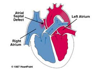
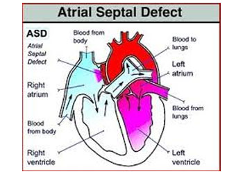
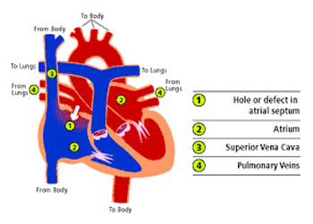
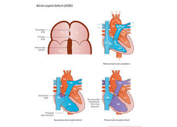
Atrial septal defect is a condition where a hole is found in the wall between the heart’s upper chambers. The defect is present from birth and in many cases, closes on own during early childhood. Hearts and lungs will be at risk of damage when the defects are large. There are no visible risks to the body when small ASDs are found. Surgery is recommended to treat this kind of defect. If the defect is not detected in adults, then it can cause heart failure and similar problems later on.


Let’s have a look at different types of atrial septal defects:-
Let’s have a look at them:-Ostium secundum atrial septal defect – It’s the most common type. In this defect, patients are found with a peculiarly large opening in the atrial septum.
Ostium primum atrial septal defect – It’s not very common defect. This defect is found in atrial septum’s the most anterior and inferior aspect.
Sinus venosus atrial septal defect – A defect involving venous inflow. It does not account more than 1% of atrial septal defect and is found in the upper part of the atrial septum.
Common or single atrium – Linked with heterotaxy syndrome. It’s a rare form of congenital defect characterized by absence or virtual absence of atrial.
Mixed atrial septal defect – Linked with the involvement of different septal zones. This defect accounts for only 7% of all atrial septal defects.
No visible signs or symptoms are found in babied born with atrial septal defects. The same happens with adult where no symptoms are found till the age of 30 or so. There are some specific signs that give the indication of the defect, like:-


Surgery is recommended by doctors to treat ASDs of medium to large sizes. However, surgical procedure is not considered in cases where patients suffer from severe pulmonary hypertension. In the surgery, the doctor would patch or plug the unusual opening between the atria. There are two procedures and doctors use either based on the condition and needs of patients.
One of procedures is cardiac catheterization where a catheter or thin tube is inserted and imaging techniques are used. The second procedure is open-heart surgery where a cut is made in the chest to close the hole. After the surgery, the follow-up care would depend on the kind and intensity of the problem.